- Titulinis
- Produktai
- Oftalmologija ir optometrija
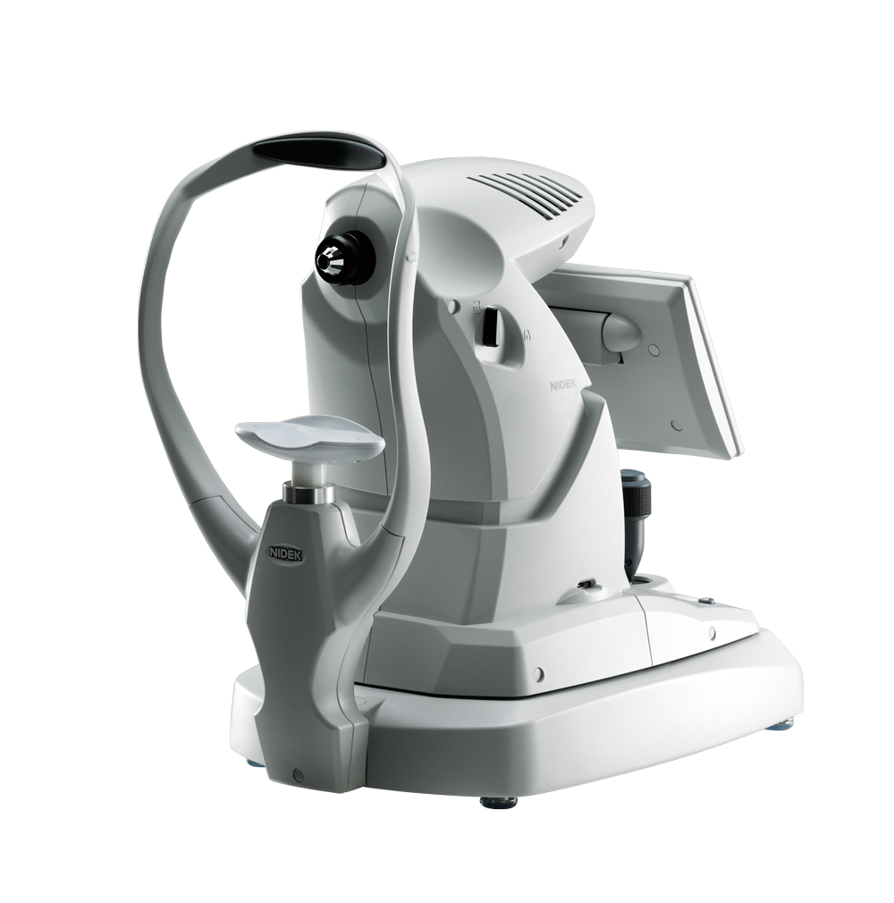
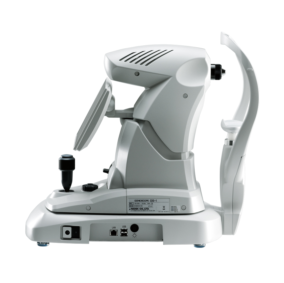
- Gonioskopas GS-1
Pirmas pasaulyje skaitmeninis gonioskopas NIDEK GS-1.
Specialus objektyvo dizainas, skirtas 360° vaizdavimui.
Galimybė saugoti ir archyvuoti paciento gonioskopinius vaizdus.
- Išmanusis „kampo aptikimas“
- Fokusavimo gylio reguliavimas
Parsisiųskite PDF brošiūrą:


Automated gonioscopy
Document. Assess. Plan.
A picture is worth a thousand words


Instant documentation with the GS-1
The GS-1 instantly documents the iridocorneal angle in real-color photographs and stores them in the device. Hence, there is no need for detailed drawing or sketches in the patient chart to record angle pathology. Photographs from the GS-1 can be attached to the patient file or chart. Documentation with the GS-1 is far simpler and much more definitive than subjective sketches of pathology.


Easy documentation and assessment with the GS-1.

The photographs can be checked on the touch screen of the device and easily magnified.
Documentation to be shared
Assessment of angle structure and pathology is easily facilitated by reviewing the digital photographs during clinical evaluation rather than performing manual gonioscopy. The digital photographs from the GS-1 can be shared from technicians to clinicians, with fellow clinicians, the referring physicians and patients, alongside complete documentation of GS-1 findings.

Focus on assessment and planning

Consultation with fellow clinicians

Effective information transfer to partner clinics

Easier patient consent
Assess and plan with documentation
The GS-1 frees up time for the clinicians to assess and plan treatment. The digital goniophotographs add the convenience of re-assessing the entire angle at any time. High resolution color photographs enhance the quality of assessment and allow comprehensive follow-up.
Postoperative examples*

MIGS

Laser iridotomy

Tube

Trabeculectomy
Progression of neovascularization (after 5 months)*

* Image courtesy of Prof C. E. TRAVERSO, MD, Clinica Oculistica, Di.N.O.G.M.I., University of Genova - Ospedale Policlinico S. Martino, Italy
GS-1 features
Automated circumferential goniophotography
An internal optical system automatically rotates and acquires color photographs of the iridocorneal angle in 16 directions / 360 degrees documenting the entire angle. Each direction can be captured in 17 different foci, enabling a versatile approach to iridocorneal angle photography.

Export images
The images acquired by the GS-1 can be displayed as a single image, with circular stitching, or linear stitching. In addition to detailed assessment with single image, stitching allows localization of features/pathologies within the entire angle. High-resolution color images are exported in JPEG, PNG and PDF files.

Non-contact gel immersion measurement
To ensure patient comfort, a coupling gel is used during image acquisition. The multimirror prism is not intended to touch the cornea.

Automated gonioscopy
Document. Assess. Plan.
A picture is worth a thousand words
Instant documentation with the GS-1
The GS-1 instantly documents the iridocorneal angle in real-color photographs and stores them in the device. Hence, there is no need for detailed drawing or sketches in the patient chart to record angle pathology. Photographs from the GS-1 can be attached to the patient file or chart. Documentation with the GS-1 is far simpler and much more definitive than subjective sketches of pathology.

Easy documentation and assessment with the GS-1.

The photographs can be checked on the touch screen of the device and easily magnified.
Documentation to be shared
Assessment of angle structure and pathology is easily facilitated by reviewing the digital photographs during clinical evaluation rather than performing manual gonioscopy. The digital photographs from the GS-1 can be shared from technicians to clinicians, with fellow clinicians, the referring physicians and patients, alongside complete documentation of GS-1 findings.
Focus on assessment and planning
Consultation with fellow clinicians
Effective information transfer to partner clinics
Easier patient consent
Assess and plan with documentation
The GS-1 frees up time for the clinicians to assess and plan treatment. The digital goniophotographs add the convenience of re-assessing the entire angle at any time. High resolution color photographs enhance the quality of assessment and allow comprehensive follow-up.
Postoperative examples*

MIGS

Laser iridotomy

Tube

Trabeculectomy
Progression of neovascularization (after 5 months)*
* Image courtesy of Prof C. E. TRAVERSO, MD, Clinica Oculistica, Di.N.O.G.M.I., University of Genova - Ospedale Policlinico S. Martino, Italy
GS-1 features
Automated circumferential goniophotography
An internal optical system automatically rotates and acquires color photographs of the iridocorneal angle in 16 directions / 360 degrees documenting the entire angle. Each direction can be captured in 17 different foci, enabling a versatile approach to iridocorneal angle photography.
Export images
The images acquired by the GS-1 can be displayed as a single image, with circular stitching, or linear stitching. In addition to detailed assessment with single image, stitching allows localization of features/pathologies within the entire angle. High-resolution color images are exported in JPEG, PNG and PDF files.
Non-contact gel immersion measurement
To ensure patient comfort, a coupling gel is used during image acquisition. The multimirror prism is not intended to touch the cornea.
NOTE