Manufacturer: NIDEK CO.,LTD. (Japan)
Warranty: 24 months.
Link to manufacturer's website: https://www.nidek-intl.com/product/ophthaloptom/diagnostic/dia_retina/duo2.html
Features:
- Fundus image acquisition with macula and disc capture in one image on OCT;
- Combined diagnosis of macular and disc pathologies;
- Denoising technique with deep learning;
- Quick acquisition of high definition B-scan images from a single-frame image;
- Fundus autofluorescence (FAF);
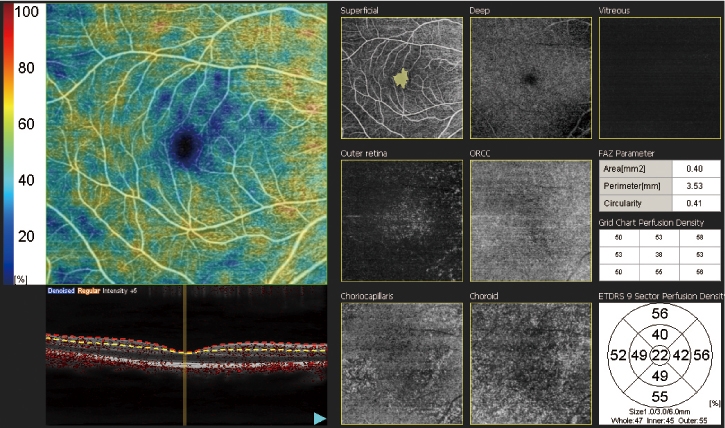
- AngioScan - optional feature.

Ideal solution for faster screening and efficient clinical workflow. Optional AngioScan is now available for OCT-Angiography imaging and diagnostics.
<<Video presentation >>

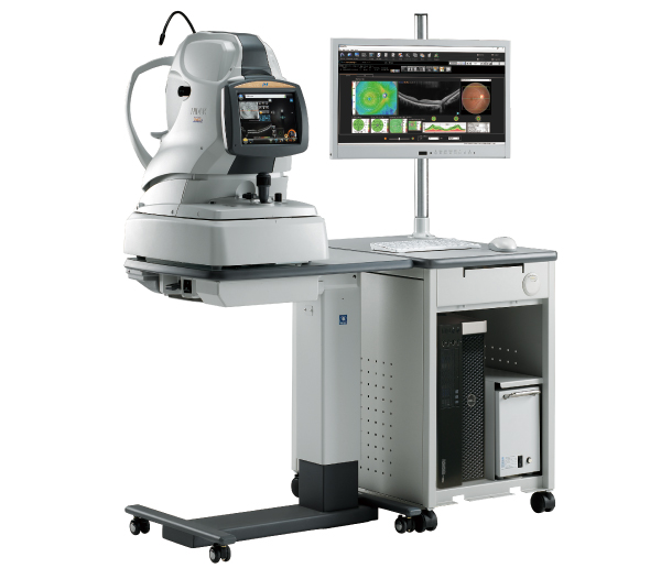


Easy operation with 3-D auto tracking, auto shot, and user friendly interface
The acclaimed 3-D auto tracking and auto shot functions allow easy imaging of the fundus and all its features. Each standard and professional mode has a different image capture interface which can be selected based on clinic preference.


Standard mode / Professional mode
High definition images
For OCT imaging, up to 50 images can be averaged and the OCT sensitivity is selectable among ultra fine, fine, and regular sensitivities based on ocular pathology. The Retina Scan Duo™ has a built-in 12-megapixel CCD camera, producing high quality fundus images.

Denoised from a single-frame image

Averaged from 50 images
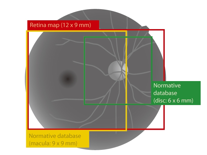
Wide area scan (12 x 9 mm) / Wide area normative database (9 x 9 mm)
A 12 x 9 mm wide area image centered on the macula can be captured with the Retina Scan Duo™. The 9 x 9 mm normative database provides a color-coded map indicating distribution range of the patient's macular thickness in a population of normal eyes.


Multiple OCT scan patterns
A wide range of scanning patterns are available to allow the practitioner to select a scan that suits the retinal region and ocular pathology.

* The anterior segment adapter is optional.
Value added features
Additional value added features include fundus autofluorescence (FAF) and En face OCT.

Color fundus image

FAF image
* The fundus autofluorescence (FAF) function is available for the FAF model.
* Photos courtesy of Kariya Toyota General Hospital.

En face OCT image
Various reports

Macula map (both eyes)

Macula line (both eyes)
-
AngioScan

-
Long axial length normative database
-
Anterior segment adapter