


Wide, deep, high-resolution imaging
With RS-1 Glauvas, a single B-scan image clearly presents the area from the optic nerve head to the temporal vascular arcade, and the 4.2 mm depth B-scan imaging readily captures the oblate retinal shape of myopic eyes. Improvements in AngioScan OCT-Angiography include wider and clearer images for assessing chorioretinal microvasculature.

Macula line 16.5 mm / Scan depth 4.2 mm

OCT-Angiography 6 x 6, 9 x 9, 12 x 12 mm
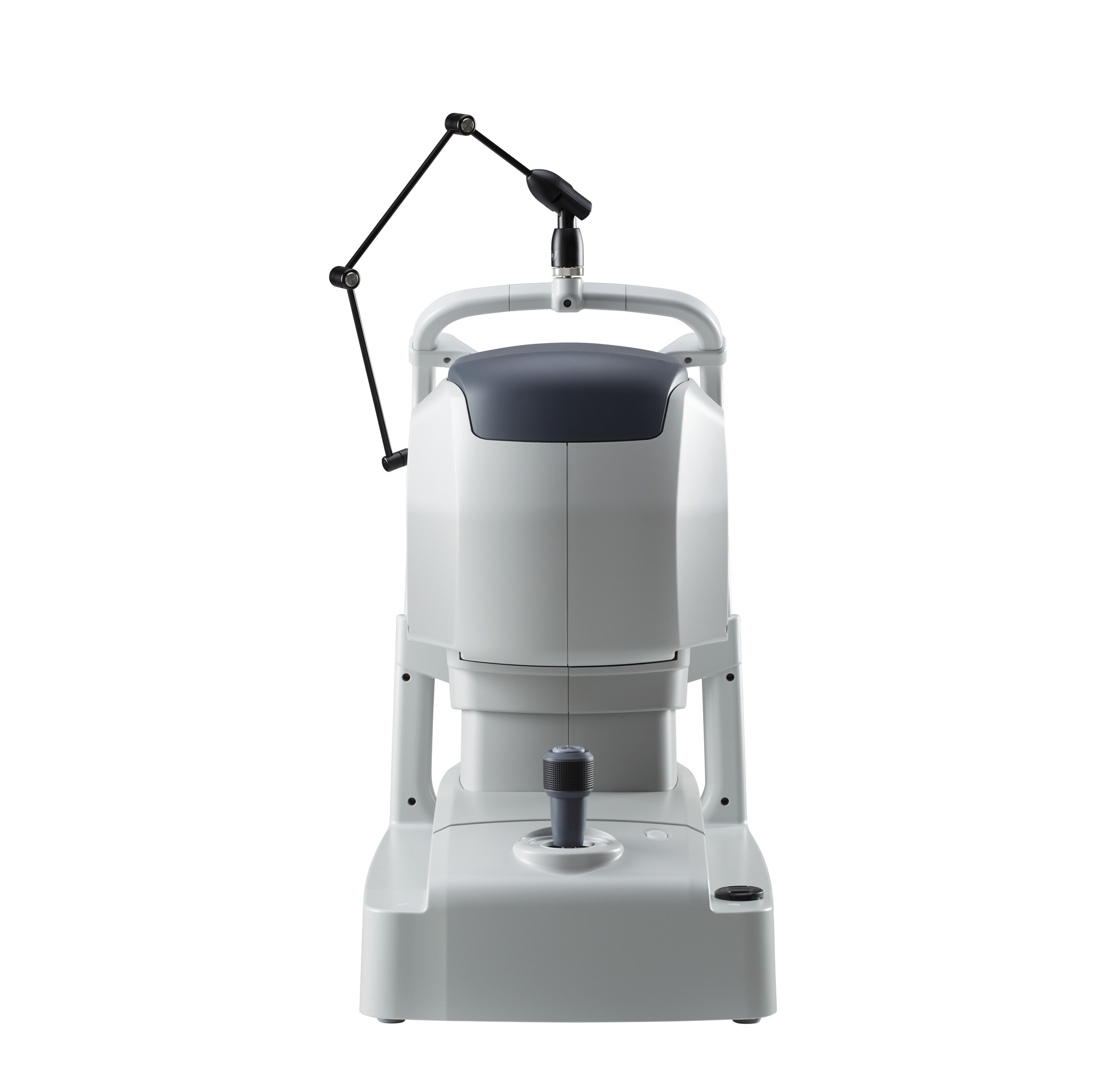
Effortless operation and interpretation
Easy image capture with automated functions
The auto alignment and auto switch functions allow anyone to effortlessly capture images. Operators need to only adjust the chinrest height and click Optimize and Capture.

Advanced analytics
Glaucoma analysis in myopia
The long axial length normative database*1 presents analysis with axial length compensation, allowing for a more accurate glaucoma assessment in patients with axial myopia. The OCT Viewer automatically switches to this database as required, by using the axial length*2 which is a parameter for scan width correction.

Sample case: Patient with 27.0 mm axial length*2
Less false positives with deep learning segmentation (DL segmentation)
The accuracy of segmentation affects the outcomes of glaucoma analysis. DL segmentation reduces artifacts and errors in the normative database and thickness maps even in eyes with opacities, thus decreasing false positives and enhancing clinic efficiency by reducing unnecessary follow-up visits. Additionally, the scan width correction allows precise analytics based on the patient’s axial length*2.

*1 Data was collected from a sample of Asian patients.
*2 The value of the axial length is obtained based on the results of the OCT image capture and differs from the actual measured value of the axial length.
Structural Normality Map (SN Map) improving diagnostic confidence on early detection
The DL segmentation provides an SN Map that presents structural abnormalities and changes. This functionality aids clinicians in detecting minute structural changes at a glance, contributing to greater diagnostic confidence even for early signs of retinal changes.

Less affected by a lesion or image contrast, the DL segmentation detects the edge of each layer on a B-scan image. Based on this highly precise segmentation, the SN Map can show subtle changes.


NOTE