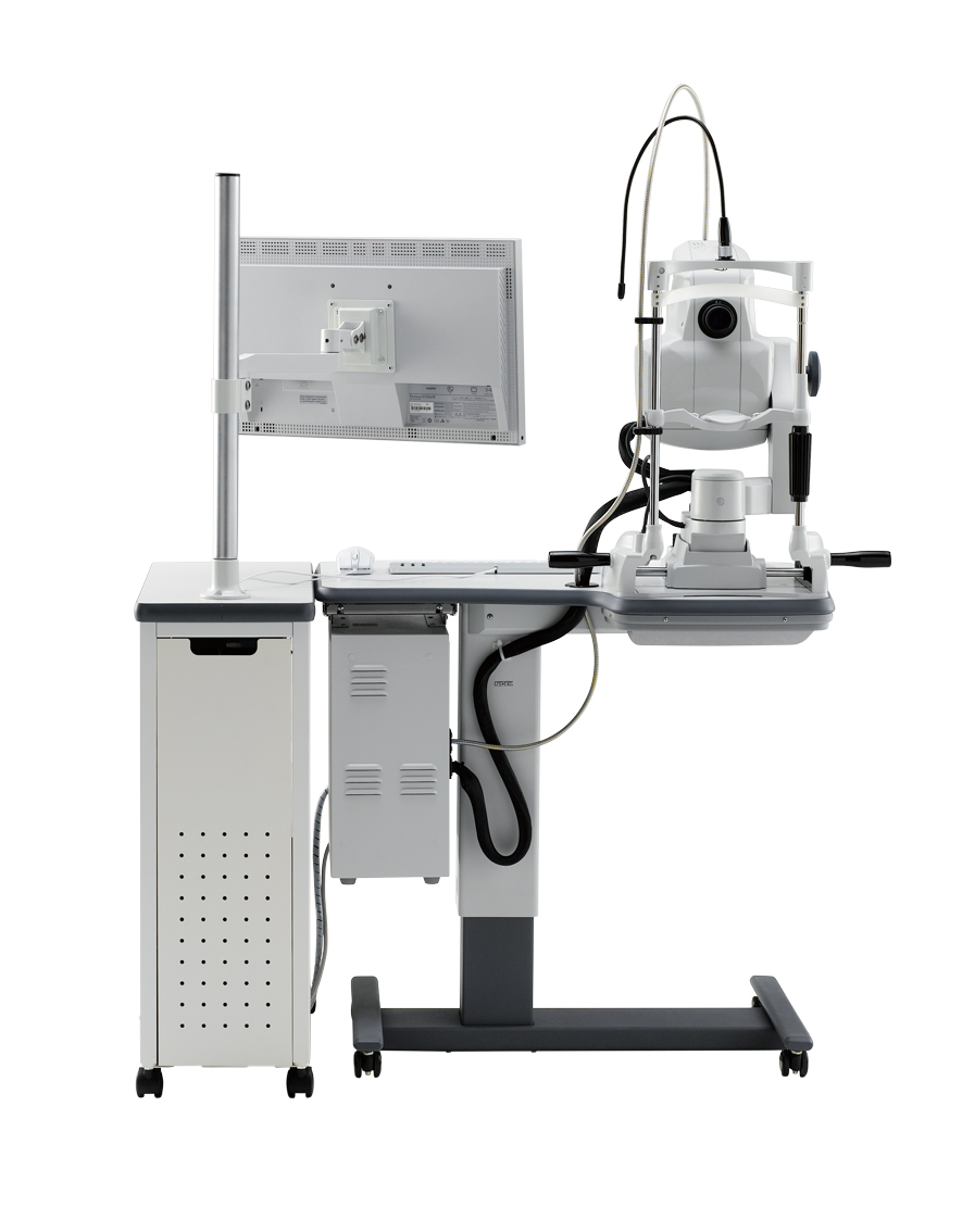
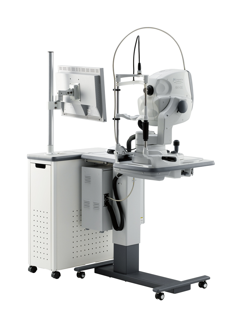
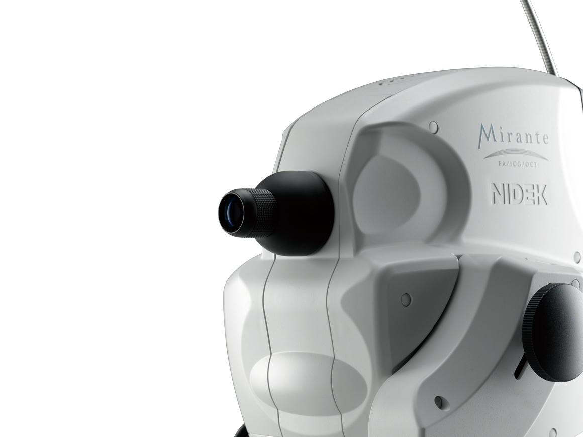
Scanning Laser Ophthalmoscope
Mirante SLO/OCT
Mirante SLO
Manufacturer: NIDEK CO.,LTD.
Warranty: 18 months.
Manufacturer's webpage: Scanning Laser Ophthalmoscope Mirante SLO/OCT Mirante SLO | Retina & Glaucoma | NIDEK CO.,LTD. (nidek-intl.com)

- The ultimate multimodal imaging platform;
For the SLO/OCT model:
- Color / FA / ICG / Blue-FAF / Green-FAF / Retro mode;
- OCT / OCT-Angiography*;
For the SLO model:
- Color / FA* / ICG* / Blue-FAF /Green-FAF / Retro mode; - Ultra wide field x ultra HD image*;
- Unsurpassed color;
- Dynamic/Simultaneous FA and ICG;
- Unique Retro mode;
- HD wide area OCT;
- Fly Through function.
*Optional


The ultimate multimodal imaging platform
For the SLO/OCT model
- Color / FA / ICG / Blue-FAF / Green-FAF / Retro mode
- OCT / OCT-Angiography*
For the SLO model
- Color / FA* / ICG* / Blue-FAF /Green-FAF / Retro mode
* Optional


Ultra wide field x ultra HD image *
163° ultra wide field image
The clear image of the entire 163° field of view** enables detailed evaluation of pathologies from the fovea to the extreme periphery.
* Ultra wide field imaging is available with the optional wide-field adapter.
** Measured from the center of the eye

Color

Retro mode

FA

ICG
Ultra 4K HD and averaging function for unparalleled clarity
4,096 x 4,096 pixel imaging captures every detail of the retina and choroid. Additionally, zooming in allows high magnification, clear visualization of subtle changes in pathology, and resolution of the fine details of capillaries.
New FlexTrack algorithm corrects image distortion due to unstable fixation and enhances averaging quality.

Distorted image due to poor fixation

Corrected image using FlexTrack
Unsurpassed color
Three separate RGB detectors simultaneously scan different depths of retina with red, green, and blue wavelengths. A color histogram is available for fine adjustment based on pathology or practitioner preference.

Color histogram adjusted similar to slit lamp view

Color histogram adjusted similar to fundus camera image

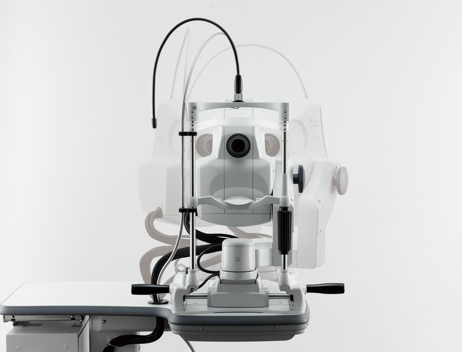
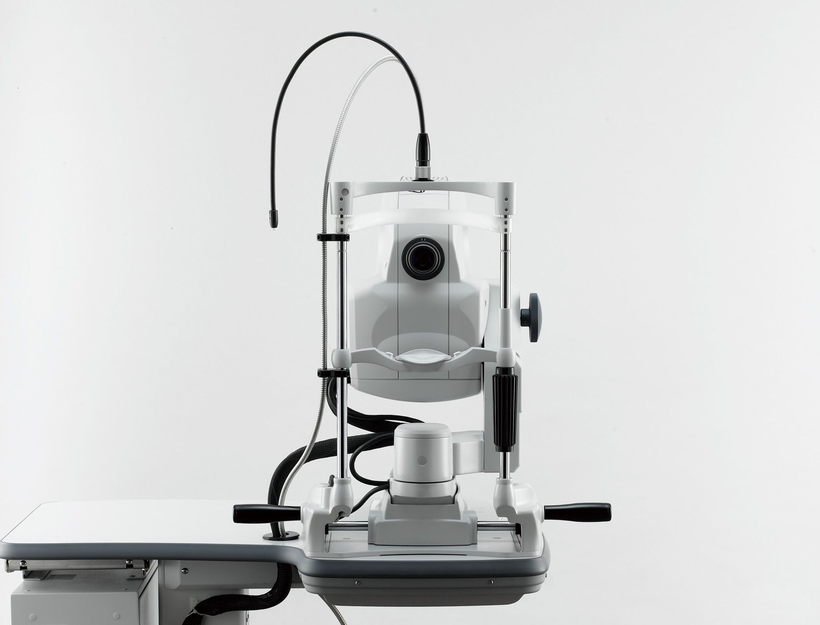
RGB detectors (Light-sensitive elements)
FA and ICG *
HD dynamic angiogram
Videos can be recorded at a maximum of 1,024 x 1,024 pixels for up to 120 seconds. Multiple short videos can be recorded during the same measurement.

Simultaneous FA and ICG
The Mirante allows simple, simultaneous acquisition of FA and ICG images.
The live IR monitoring enables alignment prior to fluorescence emission and reduces in the risk of missing the very early phase of angiography.
The Auto gain control (AGC) simultaneously adjusts contrast of each FA and ICG image, making the imaging of dynamic blood flow a very simple procedure.
* Available for the SLO/OCT model. Optional for the SLO model.

Simultaneous FA and ICG imaging display

Live IR monitoring
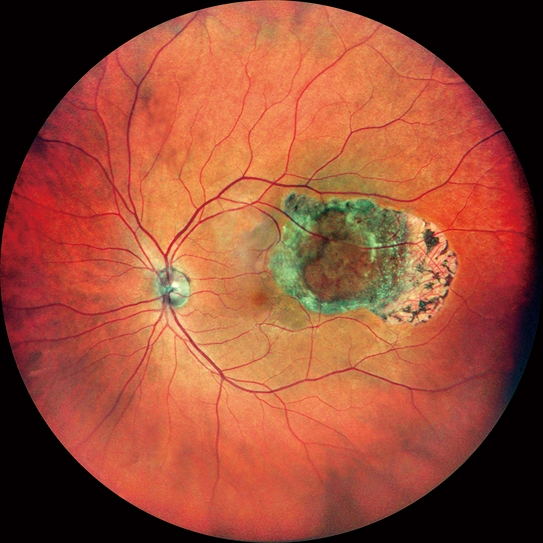
Retro mode
Retro mode is a unique non-invasive technique for detecting pathologic changes in the choroid.
This imaging modality uses scattered IR light to detect abnormal reflection in the choroid caused by drusen, edema and other subtle chorioretinal pathologies.

Color and Retro mode images (Drusen)
HD wide area OCT *
The maximum 16.5 x 12 mm area scan available with the Mirante allows wide area diagnosis including the macula and optic disc in a single shot. The ultra fine mode and tracing HD plus functions provide high quality images for detailed observation from vitreous to choroid.
* Available for the SLO/OCT model.

Macula line 16.5 mm / 2,048 A-scans

AngioScan OCT-Angiography (optional)
Fly Through function
The Fly Through function further enhances multimodal imaging by registering and synchronizing images from different modalities to view the same area while scrolling through the region of interest.

Multimodal imaging *
Various OCT modalities can be registered with microperimetry captured by NIDEK MP-3.
* Available for the SLO/OCT model.

OCT Angiography+Microperimetry

Normative DB+Microperimetry

Thickness Map DB+Microperimetry
Images courtesy
Luigi Sacco Hospital, University of Milan, Italy
Doheny Eye Center, UCLA, USA
Retina Foundation & Eye Research Center, India
Exilaser Clinic, Peru